


Volume 31, Nº 4, July and August 2018
DOI: http://www.dx.doi.org/10.5935/2359-4802.20180039
CASE REPORT
Cardiac Amyloidosis with Heart Failure and Middle Range Ejection Fraction
Antonio Jose Lagoeiro Jorge
Diane Xavier de Ávila
Enoï Guedes Vilar
Mario Luiz Ribeiro
Karima Elias Hallack Bruno
Ana Carolina Pires
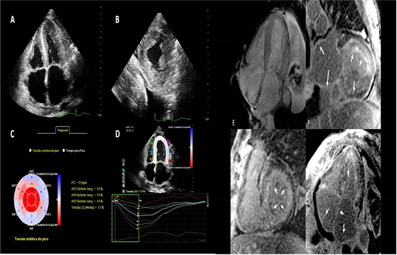
Figure 1 - (A) Apical four-chamber view showing biatrial enlargement, thickened interatrial septum, left ventricular wall hypertrophy, with hyperechoic texture of the myocardium. (B) Parasternal short axis view. Concentric left ventricular hypertrophy. (C) Bullseyes. Global longitudinal systolic strain (mean value of (-)12%), with prominent basal and medium impairment, and preserved apical mechanical function. (D) Apical four-chamber strain showing that the ratio between the apical inferior septal strain and the lower basal septal strain is greater than 3. (E) Cine-mode of the stationary state free precession showing diffuse left ventricular myocardial thickening. (F) Long axis post-gadolinium image showing diffuse late improvements (arrows) in the left atrium, as well as subendocardial enlargement of the left ventricle, more prominent in the anterior wall. (G) Short axis postgadolinium image showing diffuse subendocardial enhancement (arrows) in the left ventricle. (H) Four-chamber post-gadolinium image showing late enhancement (arrows) not only in the left chambers, but also of the right atrium.
Keywords: Heart Failure / physiopathology; Amyloidosis; Stroke Volume; Hypertrophy, Left Ventricular; Aged.