


Volume 31, Nº 2, March/April 2018
DOI: http://www.dx.doi.org/10.5935/2359-4802.20180011
REVIEW ARTICLE
Chagas Disease Cardiomyopathy
Marcus Vinicius Simões
Minna Moreira Dias Romano
André Schmidt
Káryta Suely Macedo Martins
José Antonio Marin-Neto
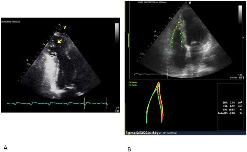
Figure 3 – Chart A: Echocardiography of apical two-chamber view of the left ventricle shows an image suggestive of a large apical aneurysm filled with thrombus (yellow arrow). Chart B: Point Tracking Technique (Speckle tracking) supporting the analysis of RV systolic function in a patient with Chagas’ disease. EDA: end-diastole area; ESA: end-systole area; FAC: Fractional area change; GLS-endo: Global longitudinal strain in the endocardial layer.
Keywords: Cardiomyopathies; Chagas Cardiomyopathy; Trypanosoma Cruzi; Chagas Disease; Heart Failure.