


Volume 113, Nº 1, July 2019
DOI: http://www.dx.doi.org/10.5935/abc.20190094
ORIGINAL ARTICLE
Characterization of Decellularized Human Pericardium for Tissue Engineering and Regenerative Medicine Applications
Luciana Wollmann
Paula Suss, João Mendonça Cesar Luzia
Andressa Schittini
George Willian Xavier da Rosa
Francisco Costa
Felipe F. Tuon
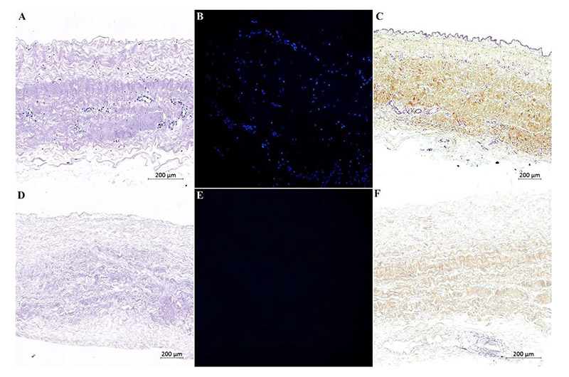
Figure 1 – Histological characterization of fresh and decellularized human pericardium. H&E and DAPI staining showed no evidence of cell in the tissue sections (D and E).H&E and RPM staining showed that decellularization did not affect the structure of collagen bundles. Howevers there was a reduction of collagen and increased thickness of the decellularized tissue compared with the fresh one (D and F).
Abstract
Background: Pericardium tissue allograft can be used for surgical repair in several procedures. One of the tissue engineering strategies is the process of decellularization. This process decreases immunogenic response, but it may modify the natural extracellular matrix composition and behavior.
Objective: The aim of this study was to evaluate the effectiveness of cell removal, maintenance of extracelular matrix properties and mechanical integrity of decellularized human pericardium using a low concentration solution of sodium dodecyl sulfate.
Methods: Decellularization was performed with sodium dodecyl sulfate and ethylenediaminetetraacetic acid. Histological analysis, DNA quantification, evaluation of glycosaminoglycans and collagen were performed. Biomechanical assay was performed using tensile test to compare the decellularization effects on tissue properties of tensile strength, elongation and elastic modulus. P < 0.05 was considered significant.
Results: There was reduction in visible nuclei present in pericardium tissue after decellularization, but it retained collagen and elastin bundles similar to fresh pericardium. The DNA contents of the decellularized pericardium were significantly reduced to less than 511.23 ± 120.4 ng per mg of dry weight (p < 0.001). The biomechanical assay showed no significant difference for fresh or decellularized tissue.
Conclusion: The decellularization process reduces cell content as well as extracellular matrix components without changing its biomechanical properties. (Arq Bras Cardiol. 2019; 113(1):11-17)
Keywords: Pericardium; Tissue Banks; Tissue Engineering/trends; Cell Separation; Glycosaminoglycans.


