


Volume 112, Nº 4, April 2019
DOI: http://www.dx.doi.org/10.5935/abc.20190064
ORIGINAL ARTICLE
Intra-Atrial Dyssynchrony Using Cardiac Magnetic Resonance to Quantify Tissue Remodeling in Patients with Atrial Fibrillation
Luisa Allen Ciuffo
João Lima
Henrique Doria de Vasconcellos
Muhammad Balouch
Susumu Tao
Saman Nazarian
David D. Spragg
Joseph E. Marine
Ronald D. Berger
Hugh Calkins
Hiroshi Ashikaga
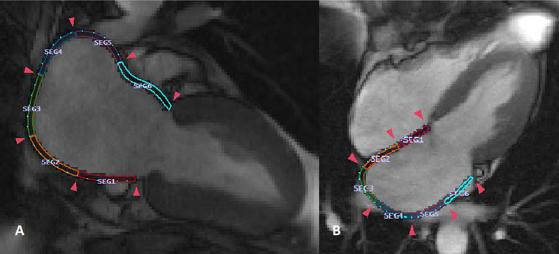
Figure 1 – Quantification of left atrial regional function using cine cardiac magnetic resonance. The figures show a total of 12 color-coded segments within the left atrium.A: Two-chamber view with six equal-length segments; B: Four-chamber view with six equal-length segments.
Abstract
Background: Recent studies suggest that left atrial (LA) late gadolinium enhancement (LGE) can quantify the underlying tissue remodeling that harbors atrial fibrillation (AF). However, quantification of LA-LGE requires labor-intensive magnetic resonance imaging acquisition and postprocessing at experienced centers. LA intra-atrial dyssynchrony assessment is na emerging imaging technique that predicts AF recurrence after catheter ablation. We hypothesized that 1) LA intra-atrial dyssynchrony is associated with LA-LGE in patients with AF and 2) LA intra-atrial dyssynchrony is greater in patients with persistent AF than in those with paroxysmal AF.
Method: We conducted a cross-sectional study comparing LA intra-atrial dyssynchrony and LA-LGE in 146 patients with a history of AF (60.0 ± 10.0 years, 30.1% nonparoxysmal AF) who underwent pre-AF ablation cardiac magnetic resonance (CMR) in sinus rhythm. Using tissue‑tracking CMR, we measured the LA longitudinal strain in two- and four-chamber views. We defined intra-atrial dyssynchrony as the standard deviation (SD) of the time to peak longitudinal strain (SD-TPS, in %) and the SD of the time to the peak pre-atrial contraction strain corrected by the cycle length (SD-TPSpreA, in %). We used the image intensity ratio (IIR) to quantify LA-LGE.
Results: Intra-atrial dyssynchrony analysis took 5 ± 9 minutes per case. Multivariable analysis showed that LA intra-atrial dyssynchrony was independently associated with LA-LGE. In addition, LA intra-atrial dyssynchrony was significantly greater in patients with persistent AF than those with paroxysmal AF. In contrast, there was no significant difference in LA-LGE between patients with persistent and paroxysmal AF. LA intra-atrial dyssynchrony showed excellent reproducibility and its analysis was less time-consuming (5 ± 9 minutes) than the LA-LGE (60 ± 20 minutes).
Conclusion: LA Intra-atrial dyssynchrony is a quick and reproducible index that is independently associated with LA-LGE to reflect the underlying tissue remodeling. (Arq Bras Cardiol. 2019; 112(4):441-450)
Keywords: Heart Atria; Atrial Fibrillation; Diagnostic Imaging; Echocardiography/methods; Magnetic Resonance Spectroscopy.


