


Volume 112, Nº 3, March 2019
DOI: http://www.dx.doi.org/10.5935/abc.20190007
ORIGINAL ARTICLE
Cardiac Evaluation in the Acute Phase of Chagas’ Disease with Post-Treatment Evolution in Patients Attended in the State of Amazonas, Brazil
Jessica Vanina Ortiz
Bruna Valessa Moutinho Pereira
Katia do Nascimento Couceiro
Monica Regina Hosannah da Silva e Silva
Susan Smith Doria
Paula Rita Leite da Silva
Edson da Fonseca de Lira
Maria das Graças Vale Barbosa Guerra
Jorge Augusto de Oliveira Guerra
João Marcos Bemfica Barbosa Ferreira
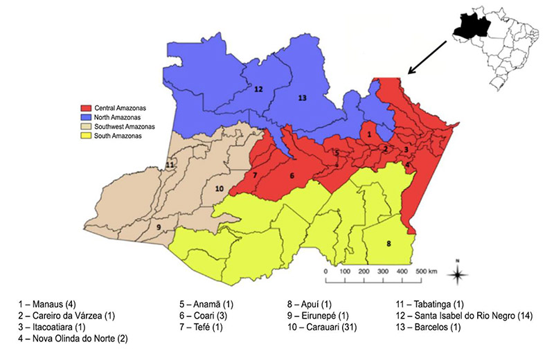
Figure 1 – Geographical distribution of the acute cases evaluated in the state of Amazonas. In parenthesis, the number of patients for each municipality.
Abstract
Background: In the past two decades, a new epidemiological profile of Chagas’ disease (CD) has been registered in the Brazilian Amazon where oral transmission has been indicated as responsible for the increase of acute cases. In the Amazonas state, five outbreaks of acute CD have been registered since 2004. The cardiac manifestations in these cases may be characterized by diffuse myocarditis, with alteration in the electrocardiogram (ECG) and transthoracic echocardiogram (TTE).
Objective: To perform a cardiac evaluation in autochthonous patients in the acute phase and at least one year after submitted to treatment for acute CD and evaluate the demographic variables associated with the presence of cardiac alterations.
Methods: We evaluated patients diagnosed with acute CD through direct parasitological or serological (IgM) methods from 2007 to 2015. These patients were treated with benznidazole and underwent ECG and TTE before and after treatment. We assumed a confidence interval of 95% (CI 95%, p < 0.05) for all variables analyzed.
Results: We observed 63 cases of an acute CD in which oral transmission corresponded to 75%. Cardiac alterations were found in 33% of the cases, with a greater frequency of ventricular repolarization alteration (13%), followed by pericardial effusion (10%) and right bundle branch block and left anterior fascicular block (2%). The follow-up occurred in 48 patients with ECG and 25 with TTE for a mean period of 15.5 ± 4.1 months after treatment. Of these, 8% presented normalization of the cardiac alterations in ECG, 62.5% remained with the normal exams. All of the patients presented normal results in TTE in the post-treatment period. As for the demographic variables, isolated cases presented more cardiac alterations than outbreaks (p = 0.044) as well as cases from Central Amazonas mesoregion (p = 0.020).
Conclusions: Although cardiac alterations have not been frequent in most of the studied population, a continuous evaluation of the clinical-epidemiological dynamics of the disease in the region is necessary in order to establish preventive measures. (Arq Bras Cardiol. 2019; 112(3):240-246)
Keywords: Chagas Disease/epidemiology; Amazoniany Ecosystem; Trypanosoma cruzi; Chagas Cardiomiopathy/physiopathology.


