


Volume 112, Nº 3, March 2019
DOI: http://www.dx.doi.org/10.5935/abc.20190042
ORIGINAL ARTICLE
Echocardiographic Correlation between Right Ventricular Function and Left Atrial Volume
Liz Andréa Villela Baroncini
Lucas José Lira Borges
Ana Cristina Camarozano
Daniela de Castro Carmo
Rubens Zenobio Darwich
Jeronimo Antonio Fortunato Junior
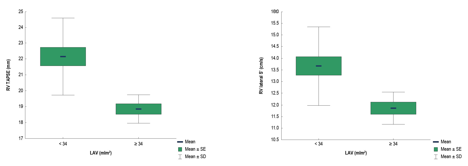
Figure 1 – Correlation between right ventricular tricuspid annular plane systolic excursion (RV TAPSE) and left atrial volume (LAV) (left panel; p < 0.001), and between lateral S’ of the right ventricle and LAV (right panel; p < 0.001). RV: right ventricular; LA: left atrium; SE: standard error; SD: standard deviation; Student’s t-test for independent samples; p < 0.05.
Abstract
Background: Few reports exist on the relationship of the left ventricular diastolic dysfunction (LVDD) with its most important features including enlargement of the left atrium and left ventricular hypertrophy (LVH), and with the right ventricular (RV) function.
Objective: To determine the correlation between the left atrial size and the RV function and dimensions in patients with and without LVDD and LVH.
Methods: Fifty patients were included, 25 (40% men) of them with LVDD, aged 67.1 ± 10.6 years (study group) and 25 without LVDD (52% men) aged 49.9 ± 16.3 years (control group). Patients underwent transthoracic echocardiography with evaluation of the left atrial size and volume (LAV), LVDD, LVH, and RV function and dimensions. P-values < 0.05 were considered statistically significant.
Results: LAV > 34 mL/m² and left atrial size > 40 mm were associated with lower absolute values of tricuspid anular plane systolic excursion (TAPSE) and RV lateral S’ (p ≤ 0.001, Pearson’s correlation coefficient -0.4 and -0.38, respectively) in the study group. Patients in the study group showed higher incidence of LVH (p = 0.02) and greater left atrial diameter (p=0.03) compared with the control group. In addition, greater left atrial diameter (p = 0.02) and LAV (p = 0.01) values were found in patients with LVDD grade II compared with LVDD grade I.
Conclusions: The present study determined, for the first time, the correlation of left atrial enlargement with progressive RV dysfunction in patients with LVDD. (Arq Bras Cardiol. 2019;112(3):249-257)
Keywords: Ventricular Dysfunction Right; Atrial Function/Physiology; Echocardiography/Methods; Blood Pressure; Heart Failure; Stroke Volume.


