


Volume 111, Nº 1, July 2018
DOI: http://www.dx.doi.org/10.5935/abc.20180091
ORIGINAL ARTICLE
Early Assessment of Right Ventricular Function in Systemic Lupus Erythematosus Patients using Strain and Strain Rate Imaging
Runlan Luo
Hongyan Cui
Dongmei Huang
Lihua Sun
Shengda Song
Mengyao Sun
Guangsen Li
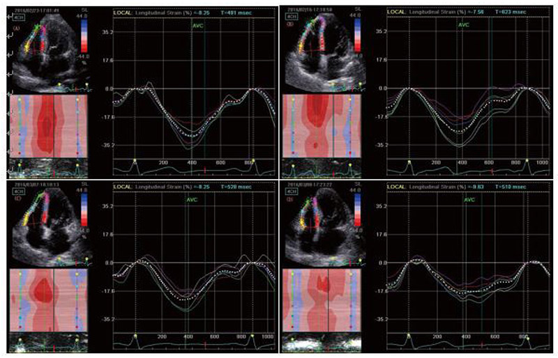
Figure 1 – Longitudinal peak systolic strain (ε) curve was obtained in right ventricular free wall for basal, middle and apical segment by 2D-STE from the apical four‑chamber view. (A) group A; (B) group B (systemic lupus erythematosus – SLE, without pulmonary hypertension); (C) group C (SLE with mild pulmonary hypertension); (D) group D (SLE with moderate-to-severe pulmonary hypertension).
Abstract
Background: Right ventricular function is a crucial factor of the prognosis of systemic lupus erythematosus (SLE).
Objectives: To evaluate the right ventricular function in SLE patients with different degrees of pulmonary hypertension (PH) by strain and strain rate imaging.
Methods: A total of 102 SLE patients and 30 healthy volunteers were studied between October 2015 and May 2016. Patients were divided into three groups according to pulmonary artery systolic pressure (PASP) estimated by echocardiography: group control (A); PASP ≤ 30 mmHg (group B, n = 37); PASP 30-50 mmHg (mild PH; group C, n = 34); and PASP ≥ 50 mmHg (moderate-to-severe PH; group D, n = 31). Longitudinal peak systolic strain (ε) and strain rate (SR), including systolic strain rate (SRs), early diastolic strain rate (SRe) and late diastolic strain rate (SRa) were measured in the basal, middle and apical segments of the right ventricular free wall in participants by two-dimensional speckle tracking echocardiography (2D-STE) from the apical four-chamber view. A p < 0.05 was set for statistical significance.
Results: The parameters of ε, SRs, SRe, and SRa were significantly decreased in groups C and D compared with groups A and B. The ε of each segments was significantly lower in group D than in group C, while there were no differences in SRs, SRe and SRa between groups C and D. Conclusions: Strain and strain rate imaging could early detect the right ventricular dysfunction in SLE patients with PH, and provide important value for clinical therapy and prognosis of these patients. (Arq Bras Cardiol. 2018; 111(1):75-81)
Keywords: Ventricular Function, Right / physiology; Lupus Erythematosus, Systemic; Hypertension, Pulmonary; Echocardiography.


