


Volume 110, Nº 5, May 2018
DOI: http://www.dx.doi.org/10.5935/abc.20180070
CASE REPORT
Surgical Epicardial CRT-D Implantation in a Patient with Complete Obstruction of the Superior Vena Cava
Gustavo Lima da Silva
Nuno Cortez-Dias
João de Sousa
Ângelo Nobre
Fausto J. Pinto
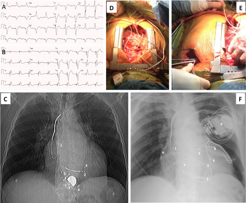
Figure 1 – A) 12-lead ECG before epicardial CRT-D implantation: sinus P waves with dissociated right ventricular epicardial pacing; B) 12-lead ECG after epicardial CRT-D implantation: sequential atrial pacing and biventricular pacing. C) Chest x ray before epicardial CRT-D implantation: 1 Abandoned endocavitary right atrial lead; 2- Abandoned endocavitary right ventricular pacing/defibrillator lead; 3- Abandoned endocavitary left ventricular lead; 4 – Epicardial mono-chamber pacemaker generator; 5 - Epicardial mono-chamber pacemaker lead. D) Intra operatory situs after lead implantation: 1 - Epicardial right atrial lead; 2 – Epicardial right ventricular outflow tract lead; 3- Left ventricular lateral lead; 4- Epicardial anterior defibrillator patch; 5- Epicardial posterior defibrillator patch. E) Intra operatory situs showing lead tunneling to left sided pre pectoral pocket. F) Chest x ray after epicardial CRT-D implantation: 1 Abandoned endocavitary right atrial lead; 2 - Abandoned endocavitary right ventricular pacing/defibrillator lead; 3 - Abandoned endocavitary left ventricular lead; 4 - Epicardial right atrial lead; 5 – Epicardial right ventricular outflow tract lead; 6- Left ventricular lateral lead; 7 - Epicardial anterior defibrillator patch; 8 - Epicardial posterior defibrillator patch; 9 – Epicardial CRT-D generator. CRT-D: cardiac resynchronization and defibrillation; ECG: eletrocardiogram.
Keywords: Heart Failure; Tachycardia, Ventricular; Vena Cava, Superior / physiopathology; Cardiac Resynchronization Therapy Devices; Cardiovascular Surgical Procedures.


