


Volume 110, Nº 3, March 2018
DOI: http://www.dx.doi.org/10.5935/abc.20180051
REVIEW ARTICLE
Practical Implications of Myocardial Viability Studies
Wilter dos Santos Ker
Thais Helena Peixoto Nunes
Marcelo Souto Nacif
Claudio Tinoco Mesquita
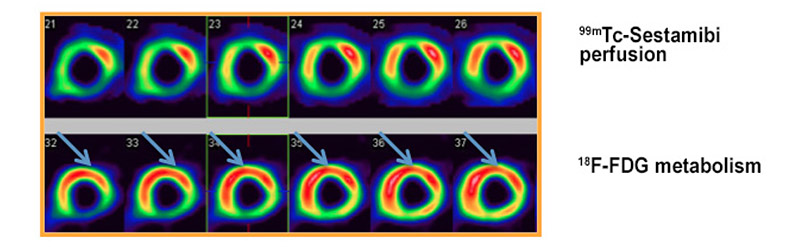
Figure 3 – Myocardial perfusion scintigraphy with 201Tálio for assessment of myocardial viability; stress imaging (upper line) and 24-hour redistribution imaging after injection of the radiotracer 201Tálio (lower line), showing improvement of perfusion in anterior (apical, medial and basal) and anterolateral (medial and basal) segments.
Abstract
Many non-invasive methods, such as imaging tests, have been developed aiming to add a contribution to existing studies in estimating patients’ prognosis after myocardial injury. This prognosis is proportional to myocardial viability, which is evaluated in coronary artery disease and left ventricular dysfunction patients only. While myocardial viability represents the likelihood of a dysfunctional muscle (resulting from decreased oxygen supply for coronary artery obstruction), hibernation represents post interventional functional recovery itself. This article proposes a review of pathophysiological basis of viability, diagnostic methods, prognosis and future perspectives of myocardial viability. An electronic bibliographic search for articles was performed in PubMed, Lilacs, Cochrane and Scielo databases, according to pre-established criteria. The studies showed the ability of many imaging techniques in detecting viable tissues in dysfunctional areas of left ventricle resulting from coronary artery injuries. These techniques can identify patients who may benefit from myocardial revascularization and indicate the most appropriate treatment.
Keywords: Tissue Survival; Diagnostic Imaging; Myocardial Revascularization / surgery; Myocardium Stunning / physiopathology.


