


Volume 110, Nº 1, January 2018
DOI: http://www.dx.doi.org/10.5935/abc.20180008
ORIGINAL ARTICLE
Melatonin-Induced Protective Effects on Cardiomyocytes Against Reperfusion Injury Partly Through Modulation of IP3R and SERCA2a Via Activation of ERK1
Shunying Hu
Pingjun Zhu
Hao Zhou
Ying Zhang
Yundai Chen
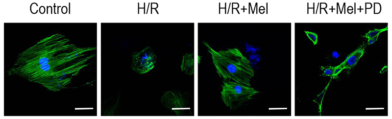
Figure 3 – Melatonin protects F-actin organization in H9C2 cells against H/R via ERK1 in vitro. Representative confocal microscopy images show H9C2 cells stained with FITC-phalloidin. The results showed that simulated H/R induced more diffuse and irregular actin disposition compared with control group. Melatonin preserved more regular and well-defined actin organization and PD98059 (ERK1 inhibitor) reduced the protection of melatonin. bar = 20μm. (Control: control group; H/R:H/R group; H/R+mel: H/R+ melatonin group; H/R+mel+PD: H/R+ melatonin+PD98059 group).
Abstract
Background: Melatonin is a neuroendocrine hormone synthesized primarily by the pineal gland that is indicated to effectively prevent myocardial reperfusion injury. It is unclear whether melatonin protects cardiac function from reperfusion injury by modulating intracellular calcium homeostasis.
Objective: Demonstrate that melatonin protect against myocardial reperfusion injury through modulating IP3R and SERCA2a to maintain calcium homeostasis via activation of ERK1 in cardiomyocytes.
Methods: In vitro experiments were performed using H9C2 cells undergoing simulative hypoxia/reoxygenation (H/R) induction. Expression level of ERK1, IP3R and SERCA2a were assessed by Western Blots. Cardiomyocytes apoptosis was detected by TUNEL. Phalloidin-staining was used to assess alteration of actin filament organization of cardiomyocytes. Fura-2 /AM was used to measure intracellular Ca2+ concentration. Performing in vivo experiments, myocardial expression of IP3R and SERCA2a were detected by immunofluorescence staining using myocardial ischemia/ reperfusion (I/R) model in rats.
Results: In vitro results showed that melatonin induces ERK1 activation in cardiomyocytes against H/R which was inhibited by PD98059 (ERK1 inhibitor). The results showed melatonin inhibit apoptosis of cardiomyocytes and improve actin filament organization in cardiomyocytes against H/R, because both could be reversed by PD98059. Melatonin was showed to reduce calcium overload, further to inhibit IP3R expression and promote SERCA2a expression via ERK1 pathway in cardiomyocytes against H/R. Melatonin induced lower IP3R and higher SERCA2a expression in myocardium that were reversed by PD98059.
Conclusion: melatonin-induced cardioprotection against reperfusion injury is at least partly through modulation of IP3R and SERCA2a to maintain intracellular calcium homeostasis via activation of ERK1. (Arq Bras Cardiol. 2018; 110(1):44-51)
Keywords: Melatonin; Myocardial Reperfusion; Cardiac Myocytes; Myocardial Infarction; Heart Failure.


