


Volume 110, Nº 1, January 2018
DOI: http://www.dx.doi.org/10.5935/abc.20170189
ORIGINAL ARTICLE
Correlation of Electrocardiographic Changes with Cardiac Magnetic Resonance Findings in Patients with Hypertrophic Cardiomyopathy
Gabriela Miana de Mattos Paixão
Horácio Eduardo Veronesi
Halsted Alarcão Gomes Pereira da Silva
José Nunes de Alencar Neto
Carolina de Paulo Maldi
Luciano de Figueiredo Aguiar Filho
Ibrahim Masciarelli Francisco Pinto
Francisco Faustino de Albuquerque Carneiro de França
Edileide de Barros Correia
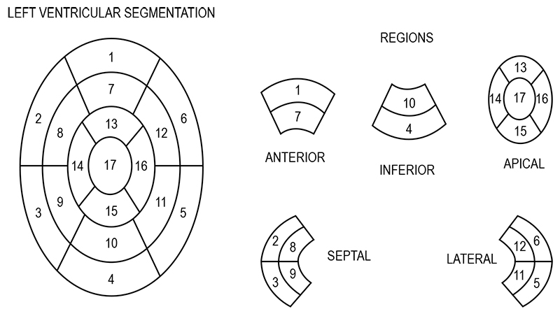
Figure 1 – Segmentação ventricular esquerda proposta pela American Heart Association
Abstract
Background: Electrocardiogram is the initial test in the investigation of heart disease. Electrocardiographic changes in hypertrophic cardiomyopathy have no set pattern, and correlates poorly with echocardiographic findings. Cardiac magnetic resonance imaging has been gaining momentum for better assessment of hypertrophy, as well as the detection of myocardial fibrosis.
Objectives: To correlate the electrocardiographic changes with the location of hypertrophy in hypertrophic cardiomyopathy by cardiac magnetic resonance.
Methods: This descriptive cross-sectional study evaluated 68 patients with confirmed diagnosis of hypertrophic cardiomyopathy by cardiac magnetic resonance. The patients’ electrocardiogram was compared with the location of the greatest myocardial hypertrophy by cardiac magnetic resonance. Statistical significance level of 5% and 95% confidence interval were adopted.
Results: Of 68 patients, 69% had septal hypertrophy, 21% concentric and 10% apical hypertrophies. Concentric hypertrophy showed the greatest myocardial fibrosis mass (p < 0.001) and the greatest R wave size in D1 (p = 0.0280). The amplitudes of R waves in V5 and V6 (p = 0.0391, p = 0.0148) were higher in apical hypertrophy, with statistical significance. Apical hypertrophy was also associated with higher T wave negativity in D1, V5 and V6 (p < 0.001). Strain pattern was found in 100% of the patients with apical hypertrophy (p < 0.001).
Conclusion: The location of myocardial hypertrophy by cardiac magnetic resonance can be correlated with electrocardiographic changes, especially for apical hypertrophy. (Arq Bras Cardiol. 2018; 110(1):52-59)
Keywords: Hypertrophic cardiomyopathy / genetic; Electrocardiography; Magnetic Resonance Spectroscopy.


