


Volume 114, Nº 1, Supplement 1, July 2020
DOI: https://doi.org/10.36660/abc.20190323
CASE REPORT
A Complicated “One Segment” Myocardial Infarction: The Role of Cardiovascular Imaging
Ana Rita Pereira
Ana Rita Almeida
Inês Cruz
Luis Rocha Lopes
Maria José Loureiro
Hélder Pereira
Video 1 – Transthoracic echocardiogram, four-chamber view, showing a non-dilated left ventricle with lateral hypokinesia and a moderate pericardial effusion with partial diastolic collapse of right cavities.
Video 2 – Ventriculography showing no apparent ventricular rupture or segmental wall motion abnormalities.
Video 3 – Cardiac magnetic resonance imaging, steady-state free precession cine images, sequentially short-axis, four-chamber and three-chamber views, showing dyskinesia of the mid-segment of the lateral wall and a saccular protuberance between the mid-segments of the lateral and inferolateral walls, suggesting a pseudoaneurysm.
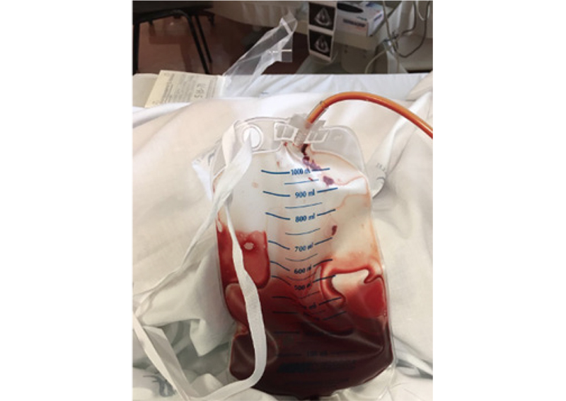
Figure 2 – Drainage system of percutaneous pericardiocentesis with around 200 mL of hematic pericardial fluid.
Keywords: Heart Rupture; Myocardial Infarction; Aneurysm,False; Diagnostic, Imaging; Echocardiography/methods.


