


Volume 114, Nº 5, May 2020
DOI: https://doi.org/10.36660/abc.20200268
CASE REPORT
Novel Coronavirus Pneumonia and Cardiomyopathy: A Case Report
Mustafa Ahmet Huyut
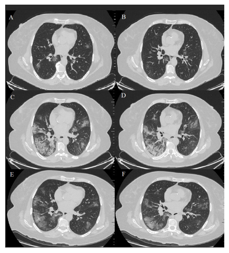
Figure 1 – Axial CT images. (A-B): At admission, CT shows the bilateral presence of mild GGOs in the parenchyma. (C-D): On day 6, a repeated CT was consistent with increasing expansion of the GGOs and progressed consolidations, which are called crazy paving consolidations. (E-F): On day 12, a repeated CT showed that the previous consolidations and GGOs in both lungs were mostly absorbed, leaving fibrous lesions that may indicate residual organizing pneumonia. CT: computer tomography, GGOs: ground-glass opacities.
Keywords: Figure 1 – Axial CT images. (A-B): At admission, CT shows the bilateral presence of mild GGOs in the parenchyma. (C-D): On day 6, a repeated CT was consistent with increasing expansion of the GGOs and progressed consolidations, which are called crazy paving consolidations. (E-F): On day 12, a repeated CT showed that the previous consolidations and GGOs in both lungs were mostly absorbed, leaving fibrous lesions that may indicate residual organizing pneumonia. CT: computer tomography, GGOs: ground-glass opacities.


