


Volume 114, Nº 4, Supplement, April 2020
DOI: https://doi.org/10.36660/abc.20190405
CASE REPORT
Recurrent Atrial Myxoma in a Patient with Carney Complex. A Case Report and Literature Review
Laura A. Cervantes-Molina
David Ramírez-Cedillo
Italo D. Masini-Aguilera
Jaime G. López-Taylor
Michel Machuca-Hernández
Dulman O. Pineda-De Paz
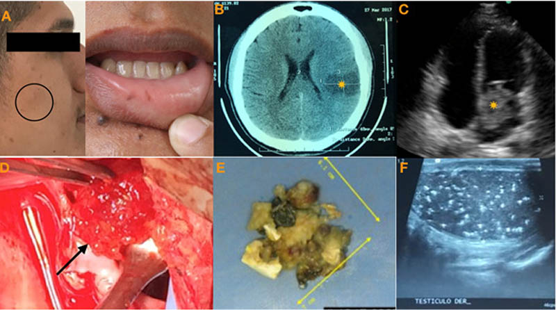
Figure 1 – Panel A. Patient’s image showing grayish nevi (arrow) and café-au-lait spots (circle). Panel B. Axial computed tomography (CT scan) of the brain showing intraaxial hypodense image in the left parieto-temporal region of 40 * 24 mm (asterisk) with mild/moderate surrounding edema. Panel C. Apical four-chamber transthoracic view showing a large left atrial mass (asterisk) and panel D showing its correlation with surgical view (arrow). Panel E. Macroscopic view of left atrial myxoma. Panel F. Right testicular ultrasound shows multiple calcifications.
Keywords: Carney Complex/complications; Atrial Myxoma; Genetic Loci; PRKAR1A.


