Volume 114, Nº 4, April 2020
DOI: https://doi.org/10.36660/abc.20190054
ORIGINAL ARTICLE
Impaired Right Ventricular Function in Heart Transplant Rejection
Luciana J. B. M. Carrion
Alice Sperotto
Raffaela Nazario
Livia A. Goldraich
Nadine Clausell
Luís Eduardo Rohde
Angela Barreto Santiago Santos

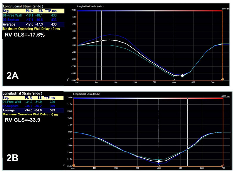
Figure 2 – Two-dimensional speckle tracking imaging for right ventricular analysis in a heart transplant recipient at the time of biopsy-proven 2R rejection (Panel 2A) and the same patient at the time of biopsy without rejection (Panel 2B). Curves represent longitudinal strain curves and the white dot represents peak-systolic strain, which were used to measure right ventricular systolic function.
Abstract
Background: The practice of screening for complications has provided high survival rates among heart transplantation (HTx) recipients.
Objectives: Our aim was to assess whether changes in left ventricular (LV) and right ventricular (RV) global longitudinal strain (GLS) are associated with cellular rejection.
Methods: Patients who underwent HTx in a single center (2015 – 2016; n = 19) were included in this retrospective analysis. A total of 170 biopsies and corresponding echocardiograms were evaluated. Comparisons were made among biopsy/echocardiogram pairs with no or mild (0R/1R) evidence of cellular rejection (n = 130 and n = 25, respectively) and those with moderate (2R) rejection episodes (n=15). P-values < 0.05 were considered statistically significant
Results: Most patients were women (58%) with 48 ± 12.4 years of age. Compared with echocardiograms from patients with 0R/1R rejection, those of patients with 2R biopsies showed greater LV posterior wall thickness, E/e’ ratio, and E/A ratio compared to the other group. LV systolic function did not differ between groups. On the other hand, RV systolic function was more reduced in the 2R group than in the other group, when evaluated by TAPSE, S wave, and RV fractional area change (all p < 0.05). Furthermore, RV GLS (−23.0 ± 4.4% in the 0R/1R group vs. −20.6 ± 4.9% in the 2R group, p = 0.038) was more reduced in the 2R group than in the 0R/1R group.
Conclusion: In HTx recipients, moderate acute cellular rejection is associated with RV systolic dysfunction as evaluated by RV strain, as well as by conventional echocardiographic parameters. Several echocardiographic parameters may be used to screen for cellular rejection. (Arq Bras Cardiol. 2020; 114(4):638-644)
Keywords: Ventricular Dysfunction, Right; Heart Transplantation; Graft Rejection; Echocardiography/methods; Strain; Speckle Tracking.


