


Volume 113, Nº 6, December 2019
DOI: http://www.dx.doi.org/10.5935/abc.20190201
ORIGINAL ARTICLE
Myocardial Perfusion by Coronary Computed Tomography in the Evaluation of Myocardial Ischemia: Simultaneous Stress Protocol with SPECT
Wilter dos Santos Ker
Daniel Gama das Neves
Tiago Augusto Magalhães
Alair Augusto Sarmet M. D. dos Santos
Claudio Tinoco Mesquita
Marcelo Souto Nacif
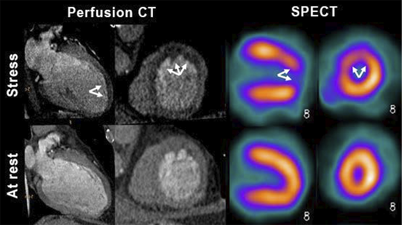
Figure 2 – Comparison between myocardial perfusion images with stress perfusion defects on computed tomography (CT) and on single-photon emission computed tomography (SPECT). Concordant example of a same patient with significant obstructive anterior descending (LAD) coronary artery disease.
Abstract
Background: Functional assessment to rule out myocardial ischemia using coronary computed tomography angiography (CCTA) is extremely important and data on the Brazilian population are still limited.
Objective: To assess the diagnostic performance of myocardial perfusion by CCTA in the detection of severe obstructive coronary artery disease (CAD) compared with single-photon emission computerized tomography (SPECT). To analyze the importance of anatomical knowledge to understand the presence of myocardial perfusion defects on SPECT imaging that is not identified on computed tomography (CT) scan.
Method: A total of 35 patients were evaluated by a simultaneous pharmacologic stress protocol. Fisher’s exact test was used to compare proportions. The patients were grouped according to the presence or absence of significant CAD. The area under the ROC curve was used to identify the diagnostic performance of CCTA and SPECT in perfusion assessment. P < 0.05 values were considered statistically significant.
Results: For detection of obstructive CAD, CT myocardial perfusion analysis yielded an area under the ROC curve of 0.84 [a 95% confidence interval (CI95%): 0.67-0.94, p < 0.001]. SPECT myocardial perfusion imaging, on the other hand, showed an AUC of 0.58 (95% CI 0.40 – 0.74, p < 0.001). In this study, false-positive results with SPECT are described.
Conclusion: Myocardial perfusion analysis by CTA displays satisfactory results compared to SPECT in the detection of obstructive CAD. CCTA can rule out false-positive results of SPECT. (Arq Bras Cardiol. 2019; 113(6):1092-1101)
Keywords: Coronary Artery Disease/physiopathology; Myocardial Ischemia; Tomography, Emission-Computed, Single‑Photon/methods; Myocardial Perfusion Imaging; Cineangiography/methods.


