


Volume 113, Nº 5, November 2019
DOI: http://www.dx.doi.org/10.5935/abc.20190155
ORIGINAL ARTICLE
3-Dimensional Echocardiography and 2-D Strain Analysis of Left Ventricular, Left Atrial and Right Ventricular Function in Healthy Brazilian Volunteers
Roberto M. Saraiva
Eliza Maria B. Scolin
Nicole P. Pacheco
Maria Eduarda Bouret
Mauro Felippe Felix Mediano
Marcelo T. Holanda
Andréa R. da Costa
Abstract
Background: New echocardiographic techniques are used in the diagnosis and prognosis of many heart diseases. However, reference values in different populations are still needed for several of these new indexes. We studied these new echocardiographic parameters in a group of Brazilians with no known cardiovascular disease.
Objective: To study values for new echocardiographic indexes in Brazilians without known cardiovascular disease and their correlation with age.
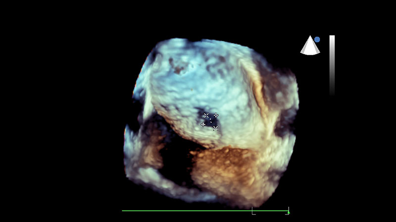
Methods: Cross-sectional study that included healthy individuals who underwent three-dimensional echocardiography (3DE) and two-dimensional speckle tracking echocardiography (STE) strain (ε) analysis. Left atrial (LA) and left ventricular (LV) function were analyzed by 3DE and STE, and right ventricular (RV) function by STE. P values < 0.05 were considered significant.
Results: Seventy-seven subjects (46.7% men; 40.4 ± 10.4 years) were included. Maximum, minimum and pre-atrial contraction (pre-A) LA volumes (ml/m2) were 21.2 ± 5.5, 7.8 ± 2.5, and 11.0 ± 3.1, respectively. Peak positive global LA ε (LAScd), peak negative global LA ε and total global LA ε (LASr) were 17.4 ± 5.2%, -13.2 ± 2.0% and 30.5 ± 5.9%, respectively. LV end-diastolic and end-systolic volumes (ml/m2) measured 57 ± 12 and 24 ± 6, and 3D LV ejection fraction measured 58 ± 6%. Global LV longitudinal, circumferential and radial ε were -19 ± 2%, -19 ± 3%, and 46 ± 12%, respectively. LV torsion measured 1.6 ± 0.70 /cm. Global longitudinal RV ε (RV-GLS) and RV free wall strain were -22 ± 3% and -24 ± 5%. Minimum LA and pre-A volumes, LV apical rotation, torsion and RV-GLS increased with age, while total and passive LA emptying fractions, LAScd, LASr, LV end-diastolic and end-systolic volumes decreased with age.
Conclusion: Values for new echocardiographic indexes in Brazilians without known cardiovascular disease and their correlation with age are presented. (Arq Bras Cardiol. 2019; 113(5):935-945)
Keywords: Cardiovascular Diseases; Echocardiography, Three-Dimensional; Reference Values; Left Ventricular Function; Right Ventricular Function; Strain; Speckle Tracking.


