


Volume 112, Nº 6, June 2019
DOI: http://www.dx.doi.org/10.5935/abc.20190105
ANATOMOPATHOLOGICAL CORRELATION
Case 3/2019 – Young Male with Intense Dyspnea, Pulmonary Infiltrate, Normal Cardiac Area and Obliteration of the Apical Portion of the Left Ventricle
Victor Sarli Issa
Luiz Alberto Benvenuti
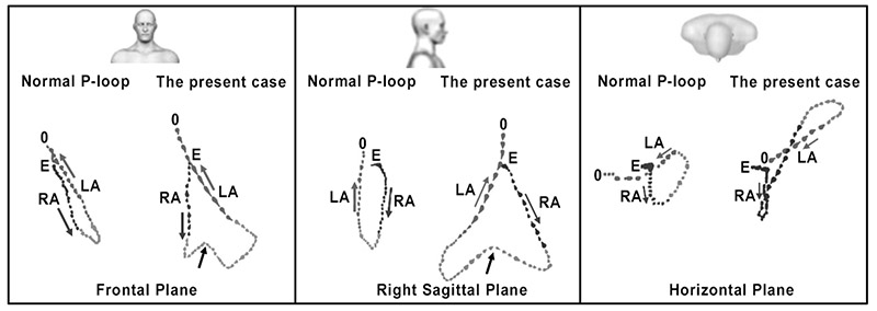
Figure 2 – Comparison between normal P loops and the present case. Frontal plane: In the present case, the maximal vector voltage is > 0.2 mV, and the morphology is broad with a notch in the middle portion (arrows). Right sagittal plane: The maximal anterior forces are ≥ 0.06 mV and maximal posterior forces are > 0.04 mV: biatrial enlargement. Horizontal plane: the normal P loop maximal vector location is located between +50° and -45°, maximal vector voltage is < 0.1 mV, maximal anterior forces are up to 0.06 mV and maximal posterior forces are up to 0.04 mV. In the present case, anterior and posterior forces exceed these values. Conclusion: biatrial enlargement and suspicion of AI by notched P loop in the frontal and right sagittal plane. RA: right atrium; LA: left atrium.
Keywords: Myocardial Infarction/physiopathology; P Wave; Diagnosis Imaging; Arrhythmias, Cardiac; Risk Factors; Percutaneous Coronary Intervention; Drug-Eluting Stents.


