


Volume 111, Nº 6, December 2018
DOI: http://www.dx.doi.org/10.5935/abc.20180218
BRIEF COMMUNICATION
Cardiovascular Manifestations of Erdheim-Chester’s Disease: A Case Series
Isabela Bispo Santos da Silva Costa
André Neder Ramires Abdo
Cristina Salvadori Bittar
Silvia Moulin Ribeiro Fonseca
Aline Sabrina Holanda Teixeira Moraes
Roberto Kalil Filho
Juliana Pereira
Ludhmila Abrahão Hajjar
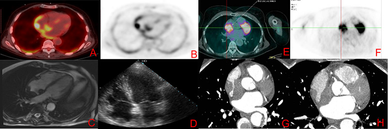
Figure 1 – Images A to D refer to case 1 and the images from E to H refer to case 2. Images A and B represent images of 18-FDG PET-CT showing lesion in the right atrium roof. The C image represent CMR image, SSFP cine 4 chambers with hypointense lesion in the right atrium roof. The D image represents a transthoracic echocardiogram image with the same topography. The images E and F represent 18-FDG PET-CT with capturing lesion in the right atrium and G and H images represent contrast computed tomography showing evidence of expansive right atrial.
Keywords: Erdheim-Chester Disease/diagnosis; Erdheim-Chester Disease/drug therapy; Erdheim-Chester Disease/pathology; Biopsy; Prognosis


