


Volume 111, Nº 5, November 2018
DOI: http://www.dx.doi.org/10.5935/abc.20180164
ORIGINAL ARTICLE
Usefulness of Preoperative Venography in Patients with Cardiac Implantable Electronic Devices Submitted to Lead Replacement or Device Upgrade Procedures
Caio Marcos de Moraes Albertini
Katia Regina da Silva
Joaquim Maurício da Motta Leal Filho
Elizabeth Sartori Crevelari
Martino Martinelli Filho
Francisco Cesar Carnevale
Roberto Costa
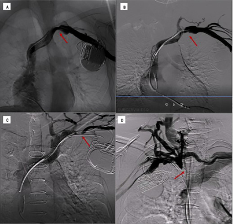
Figure 3 – Classification of venous lesions and collateral circulation. Examples of the four types of lesion according to the classification adopted in the study. Figure 3A: non-significant lesions characterized with obstruction of less than 50% of the blood vessel lumen and absence of collateral circulation; Figure 3B: moderate lesion in 51% to 70% of the vessel, with discrete collateral circulation; Figure 3C: severe lesion compromising 71% to 99% of the vessel with moderate collateral circulation; Figure 3D: venous occlusion with accentuated collateral circulation.
Abstract
Background: Venous obstructions are common in patients with transvenous cardiac implantable electronic devices, but they rarely cause immediate clinical problems. The main consequence of these lesions is the difficulty in obtaining venous access for additional leads implantation.
Objectives: We aimed to assess the prevalence and predictor factors of venous lesions in patients referred to lead reoperations, and to define the role of preoperative venography in the planning of these procedures.
Methods: From April 2013 to July 2016, contrast venography was performed in 100 patients referred to device upgrade, revision and lead extraction. Venous lesions were classified as non-significant (< 50%), moderate stenosis (51-70%), severe stenosis (71-99%) or occlusion (100%). Collateral circulation was classified as absent, discrete, moderate or accentuated. The surgical strategy was defined according to the result of the preoperative venography. Univariate analysis was used to investigate predictor factors related to the occurrence of these lesions, with 5% of significance level.
Results: Moderate venous stenosis was observed in 23%, severe in 13% and occlusions in 11%. There were no significant differences in relation to the device side or the venous segment. The usefulness of the preoperative venography to define the operative tactic was proven, and in 99% of the cases, the established surgical strategy could be performed according to plan.
Conclusions: The prevalence of venous obstruction is high in CIED recipients referred to reoperations. Venography is highly indicated as a preoperative examination for allowing the adequate surgical planning of procedures involving previous transvenous leads. (Arq Bras Cardiol. 2018; 111(5):686-696)
Keywords: Pacemaker, implantable defibrillators, phlebography, venous stenosis, extraction of leads, risk factors.


