


Volume 111, Nº 5, November 2018
DOI: http://www.dx.doi.org/10.5935/abc.20180219
CLINICORADIOLOGICAL CORRELATION
Case 6 / 2018 - Percutaneous Occlusion of a Large Ductus Arteriosus in a Low Weight Infant, with Immediate Clinical and Radiographic Improvement
Pablo Tomé Teixeirense
Vanessa de Moraes Sousa
João Felipe Barros de Toledo
Luiz Antonio Gubolino
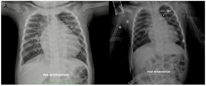
Figure 1 – A) Pre-intervention chest x-ray. There is an overall increase in the cardiac silhouette, with prominence of the right atrium, left ventricle and vascular pedicle, in addition to the pulmonary vascular network. B) Chest X-ray approximately 8h after occlusion of the defect, showing the significant decrease in the cardiac volume, notably in the right atrium and the vascular pedicle, as well as a decrease in the pulmonary vascular network.
Keywords: Infant; Down Syndrome; Heart Defects, Congenital/ surgery; Ductus Arteriosus Patent/surgery.


