Volume 111, Nº 3, September 2018
DOI: http://www.dx.doi.org/10.5935/abc.20180185
IMAGE
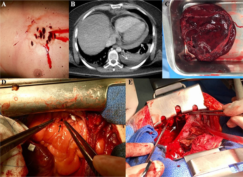
Right Ventricular Wound And Complete Mammary Artery Transection
Gregorio Laguna
Miriam Blanco
Cristina García-Rico
Yolanda Carrascal

Figure 1 – Panel A: Eleven knife-wounds localized in the left-anterior chest wall. Panel B: Axial computed tomography showed severe pericardial and left pleural effusion (white arrows). Panel C: The clot drained from the pericardial cavity. Panel D: Right ventricular perforation repaired using a monofilament suture (black arrow). Panel E: Complete mammary artery transection bleeding into left pleural cavity (white arrow).
Keywords: Stab Wounds/heart; Suicide, Attempted; Heart Injuries/cirurgia.


