


Volume 111, Nº 2, August 2018
DOI: http://www.dx.doi.org/10.5935/abc.20180110
IMAGE
Unexpected Mass in the Left Atrium
Tatiana Guimarães
Rui Plácido
Ana Catarina Quadros
José Marques da Costa
Fausto J. Pinto
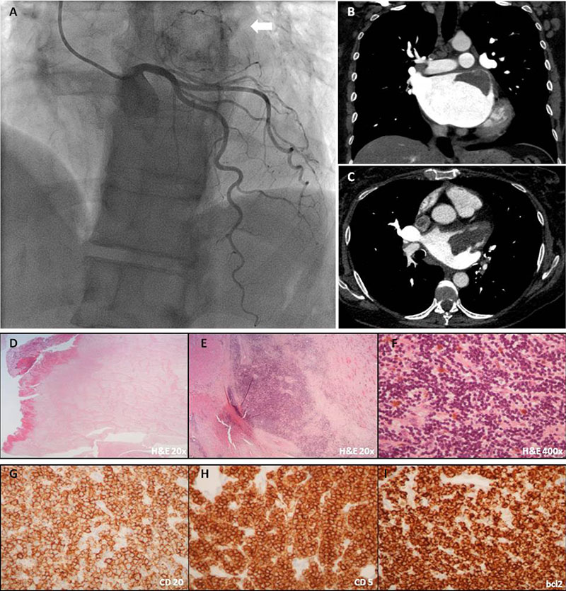
Figure 1 – (Panel A) Selective left coronary angiogram (left anterior oblique 30° position) showing an abnormal vascularized mass (arrow) in the left atrium. (Panel B and C) Coronal and axial angio-CT planes in arterial phase, respectively, demonstrating a well-delimited, homogeneous and slight hyperdense mass, along the lateral portion of the atrial roof. (Panel D) Recent thrombus, partially in organization (H&E 20x). (E and F) Myocardium and adipose tissue infiltrated by small lymphoid cells, with scant cytoplasm and nuclei with peripherally clumped chromatin (H&E 20x and 400x). (Panel G-I) CD20, CD5 and bcl2 immunoreactivity (400x), respectively.
Keywords: Heart Atria Heart Neoplasms/surgery; Leukemia,Lymphoid/ physiopathology; Mitral Valve Stenosis; Echocardiography; Coronary Angiography.


