


Volume 111, Nº 1, July 2018
DOI: http://www.dx.doi.org/10.5935/abc.20180106
IMAGE
Challenging Evaluation of Aortic Regurgitation: More Than a Quadricuspid Valve
Gonçalo Pestana
Carla Sousa
Teresa Pinho
Sara Maia
M. Júlia Maciel
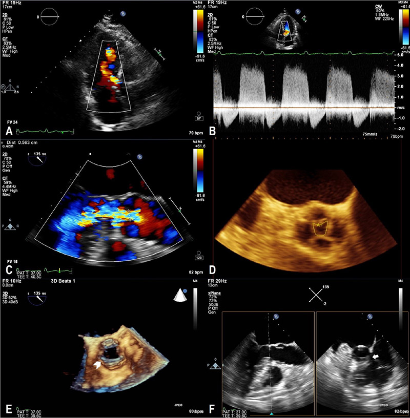
Figure 1 – Aortic regurgitation jet visualized by transthoracic echocardiogram with color Doppler (A) and its respective spectrum on continuous Doppler wave (B); large jet visualized by transesophageal echocardiogram, with a 6 mm vena contracta (C), originating from the central coaptation defect of the quadricuspid aortic valve, with a regurgitant orifice of 0.35 cm2 in three-dimensional planimetry (D); Almost circumferential thickening of the left ventricular outflow tract readily identified in the three‑dimensional image in systole (E), confirming the presence of a subaortic membrane with evaluation of orthogonal planes (F).
Keywords: Aortic Valve Insufficiency; Echocardiography, Transesophageal; Echocardiography, Three-Dimensional; Pulmonary Disease, Chronic Obstructive.


