


Volume 110, Nº 5, May 2018
DOI: http://www.dx.doi.org/10.5935/abc.20180064
IMAGE
Computed Tomography-Guided Core Needle Biopsy of Cardiac Angiosarcoma
Luis Gorospe
Alberto Cabañero-Sánchez
Gemma María Muñoz-Molina
Ana María Ayala-Carbonero
María Ángeles Fernández-Méndez
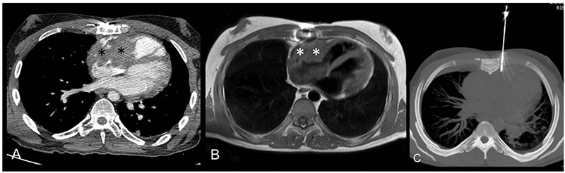
Figure 1 – A) Axial contrast-enhanced CT image showing heterogeneous mass (asterisks) infiltrating the right atrium, the right atrioventricular groove, and the right ventricle; B) Axial T1-weighted MR cardiac image showing mass (asterisks) infiltrating the right cardiac chambers; C) Axial unenhanced CT MIP (maximum intensity projection) image showing core-needle biopsy, with the tip entering the cardiac mass.
Keywords: Hemangiosarcoma/diagnosis; Hemangiosarcoma/ pathology; Biopsy, Large-Core Needle; Tomography, X-Ray Computed; Neoplasm Staging.


