


Volume 110, Nº 4, April 2018
DOI: http://www.dx.doi.org/10.5935/abc.20180057
IMAGE
Simultaneous Dual Coronary Fistulas
Ioannis Ntalas
John B. Chambers
Júlia Karády
Ronak Rajani
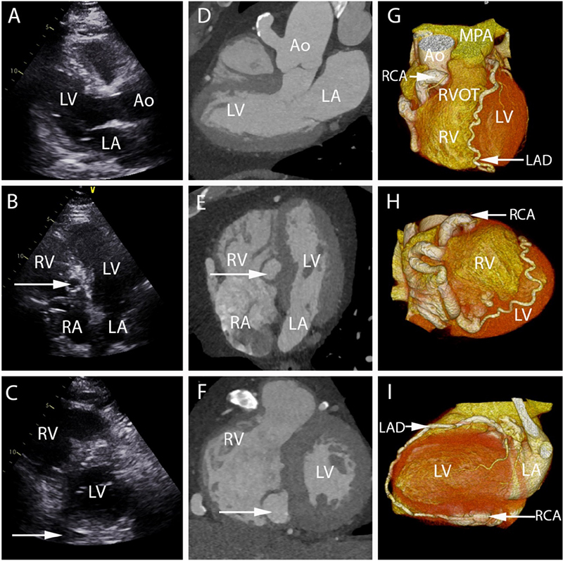
Figure 1 – A-C) show the parasternal long axis (1A), apical 4-chamber (1B) and short axis (1C) TTE views of the left ventricle. The white arrow shows the presence of a spherical structure in the 4-chamber view and a dilated blood vessel in the short axis view. D-F) show the corresponding CTCA appearances of these findings in the same “echocardiographic views”. G) shows the 3D volume rendered image of the heart with dilated and tortuous RCA and LAD. H) shows the anatomical connection of the RCA fistula to the base of the inferior RV and a continuation of the PDA to the LAD and I) shows the LAD to PDA continuation. LV: left ventricle; RV: right ventricle; LA: left atrium; RA: right atrium; Ao: aorta; MPA: main pulmonary artery; RVOT: right ventricular outflow tract; TTE: transthoracic echocardiogram; CTCA: cardiac computed tomography angiography; RCA: right coronary artery; LAD: left anterior descending artery; PDA: posterior descending artery.
Keywords: Systolic Murmurs; Echocardiography; Computed Tomography Angiography.


