


Volume 110, Nº 4, April 2018
DOI: http://www.dx.doi.org/10.5935/abc.20180065
ANATOMOPATHOLOGICAL CORRELATION
Case 2/2018 - 73-Year-Old Male with Ischemic Cardiomyopathy, Cachexia and Shock
Rafael Amorim Belo Nunes
Jussara de Almeida Bruno
Hilda Sara Monteiro Ramirez
Léa Maria Macruz Ferreira Demarchi
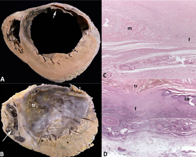
Figure 2 – A and B: Cross sections of the ventricles in the middle-basal and apical regions, respectively. Healed transmural infarction in the left ventricular anterior and lateral walls and in the anterior two-thirds of the ventricular septum, with aneurysmal formation. Myocardial hypertrophy of the left ventricular walls not affected by the infarction is evident. Organizing thrombus in the endocardium of the left ventricular anterior and septal walls (arrow) in the middle-basal region, extending to the apical region (tr). Organizing thrombus in the right ventricular endocardium of the apex (arrow). C and D: Histological sections of the left ventricular aneurysm showing an isolated group of cardiomyocytes (m) and focal calcification (ca) amid mural fibrosis (f). Hematoxylin-eosin, 25x.
Keywords: Atherosclerosis; Heart Failure/physiopathology; Cardiomyopathy, Dilated/complications; Weight Loss; Cachexia.


