Volume 110, Nº 3, March 2018
DOI: http://www.dx.doi.org/10.5935/abc.20180028
IMAGE
Partial Prosthetic Mitral Valve Dehiscence: Transapical Percutaneous Closure
Catarina Ruivo
José Ribeiro
Alberto Rodrigues
Luís Vouga
Vasco Gama

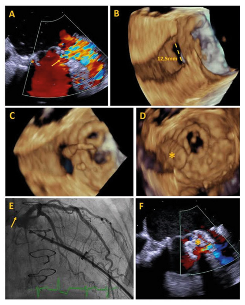
Figure 1 – Panel A: 2D peri-processual transesophageal echocardiography (TEE) shows paravalvular regurgitation (yellow arrow) between left ventricle and left atrial appendage; Panel B: Defect 3D TEE with diameter measurement; Panel C: 3D TEE guiding the guidewire through the defect; Panel D: 3D TEE showing the device (asterisk) through the defect; Panel E: left coronary angiography without vascular involvement after occlusal implant (yellow arrow); Panel F: Light residual flux detected after device deployment (asterisk).
Keywords: Endocarditis; Mitral Valve Insufficiency; Aortic Valve Insufficiency; Echocardiography, Transesophageal.


