


Volume 110, Nº 2, February 2018
DOI: http://www.dx.doi.org/10.5935/abc.20180022
CASE REPORT
A Prenatal Case of Arrhythmogenic Right Ventricular Dysplasia
Lilian Maria Lopes
Juliana Torres Pacheco
Regina Schultz
Rossana Pulcineli Vieira Francisco
Marcelo Zugaib
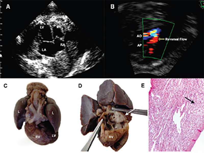
Figure 1 – Fetal echocardiography and anatomic features observed at the autopsy. (a) Four-chamber view at 36 weeks showing cardiac enlargement and left atrial dilatation. (b) Three-vessel view showing reversal flow at the ductus arteriosus level (arrow). (c) Heart and lungs with pale, enlarged right ventricle. (d) Right ventricular wall is thin and almost devoid of muscle fibers. (e) A hematoxylin-eosin stain demonstrating absence of myocardial fibers and fibrofatty tissue replacement of the anterior free wall of the right ventricle. RA: right atrium; RV: right ventricle; LA: left atrium; LV: left ventricle; L: lungs.
Keywords: Arrhythmogenic Right Ventricular Dysplasia; Fetus / echocardiography; Prenatal Care; Pregnancy.


