


Volume 110, Nº 1, January 2018
DOI: http://www.dx.doi.org/10.5935/abc.20170186
CLINICORADIOLOGICAL CORRELATION
Case 1/2018 – Preponderant Left Ventricular Restrictive Syndrome in a 28-Year-Old Woman
Edmar Atik
Danielle Haddad Syllos Dezen
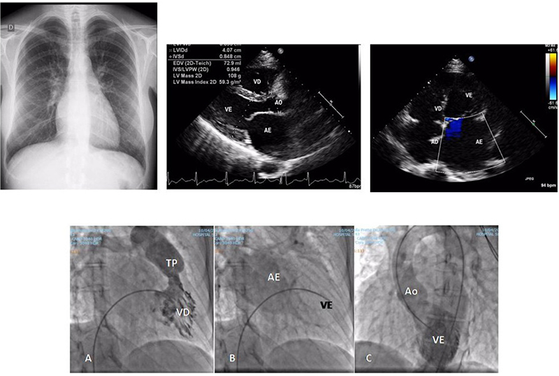
Figure 1 – Chest X-ray in AP emphasizes normal cardiac area with a clear increase of the left atrium size in a double contour in the lower right arch and slightly congested pulmonary vascular weave in the upper fields. The echocardiographic images highlight the left atrial enlargement in longitudinal and 4-chamber projection. Angiocardiograms below show right ventricular hypertrophy (A), left atrial emptying delay (B) compatible with left ventricular restrictive syndrome and this cavity with smooth and normal-sized internal borders (C).
Keywords: Cardiomyopathy, Restrictive; Atrial Fibrillation; Electroshock; Ventricular Dysfunction Left; Hypertension, Pulmonary.


